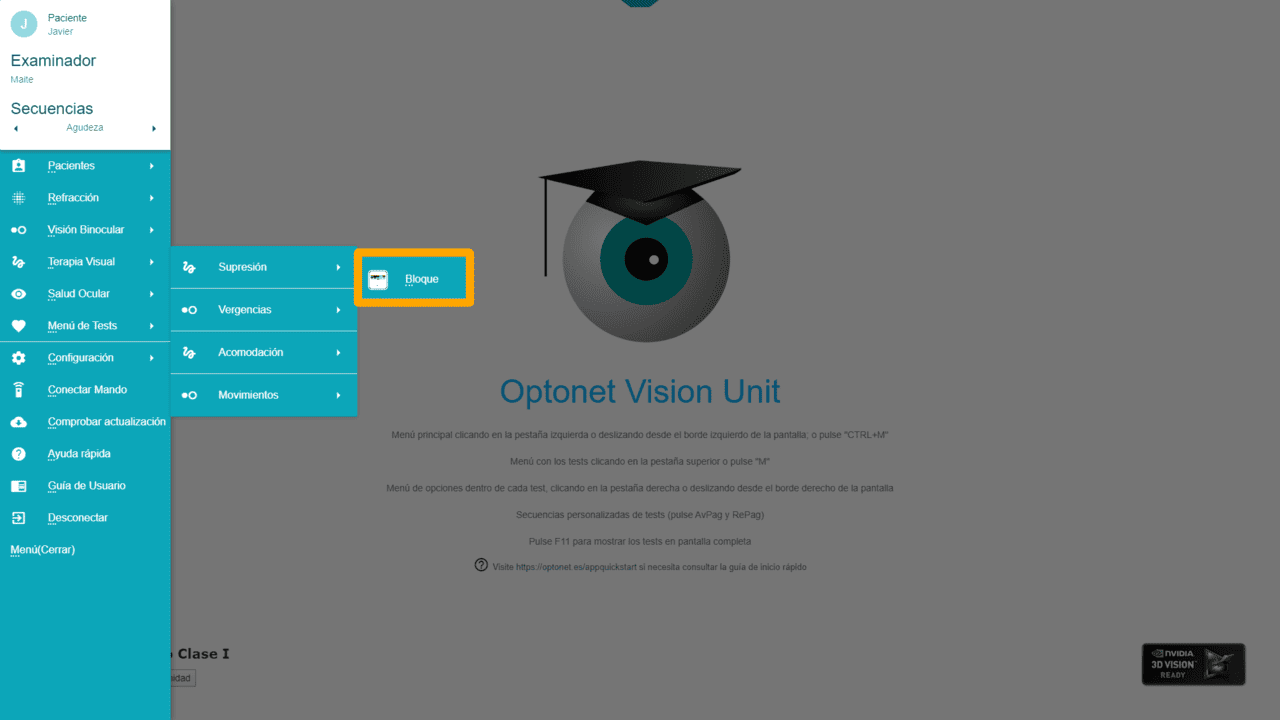
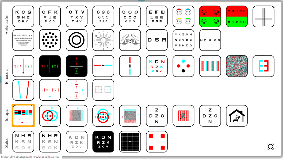
Now let’s look at each of the visual rehabilitation functions and exercises offered by the Optonet Unit. They can be accessed from the Vision Training section of the main menu or directly from the top chart menu.
Suppression and Binocular Coordination – Block Game #
Description #
This test consists of a wall with coloured blocks that must be viewed with red/blue glasses (or polarizing filters for 3D screens). With the filters, some blocks are seen only with the right eye, others only with the left, and some with both eyes; these serve as stimuli for binocular vision.
At the bottom, there is a horizontal rectangle that acts as a racket, along with a ball that makes the blocks disappear when it hits them. The racket moves from left to right when moving the mouse, and the task is to place the racket under the ball to return it against the wall. The goal is to eliminate the maximum number of blocks without missing the ball.
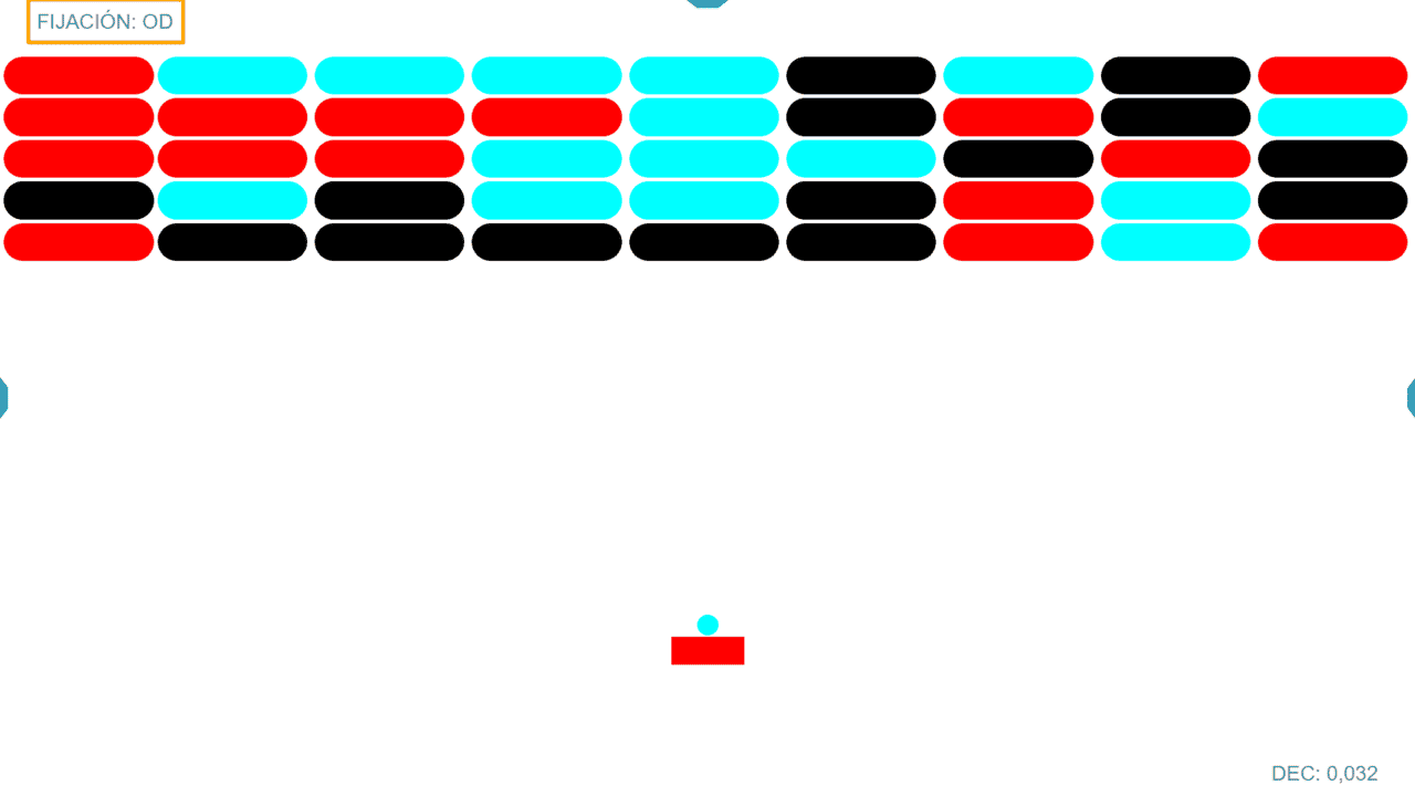
Through the filters, the racket is seen only with one eye and the ball with the other eye. At the top left of the screen, it indicates which eye sees the moving ball (the fixing eye), (the red filter always in front of the right eye). The size of the racket is displayed in the lower right corner in the different AV notations included in the program.
For the patient to return the ball with the racket, they must use and coordinate both eyes. Therefore, this exercise can be used as anti-suppression therapy and also to improve binocular coordination. If the patient suppresses one eye or cannot control their heterophoria, they will have difficulty in this game.
Menu #
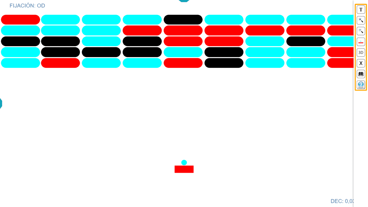
Clicking on the tab to the right of the screen (or swiping left on touch screens) opens the tools menu with various options. Hovering the mouse over each button indicates what that button is for, and also shows the keyboard letter that performs the same function (in brackets). Below, we explain the buttons.
Training #
The first Therapy button is used to define the exercise. A window opens where we choose the size of the racket, the speed of the ball, and the time we want the exercise to last. It is always recommended to start with an easy exercise and gradually increase the difficulty (e.g., start with a large racket, slow speed, and short time).
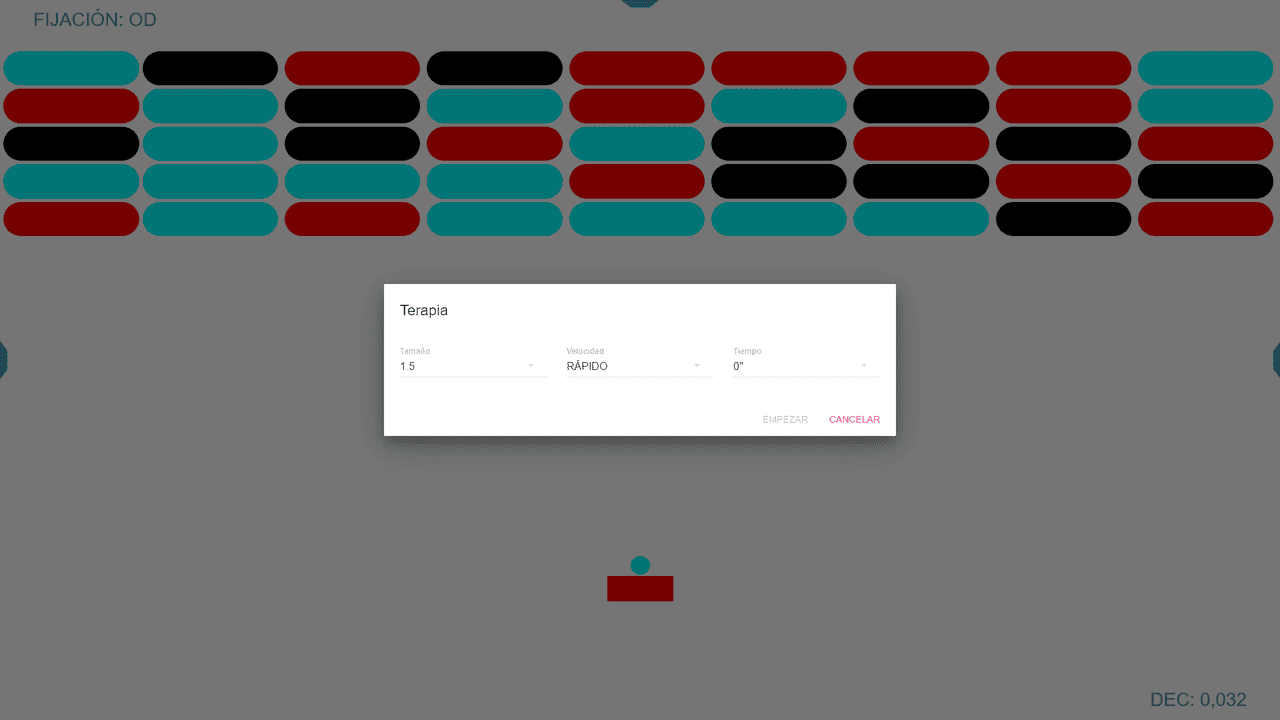
The size of the racket is expressed in log AV units. The smaller the size of the racket, the more difficult the activity is, as it requires greater precision in binocular alignment and eye-hand coordination.
The speed of the ball has three options: fast, normal, and slow (it’s more difficult the faster it is).
The time the exercise should last. It must always be indicated before starting, from 30 seconds to 5 minutes (in 30-second steps).
Once the parameters are set, we press the “Start” button to begin the activity.
The size of the racket can also be modified with the zoom buttons, or by pressing the + and – keys. The AV value corresponding to the racket is displayed in the lower right corner of the screen in the chosen notation; but by clicking on that number (or pressing the “W” key on the keyboard) we can switch to any of the other AV notations allowed by the Unit.
Colour Distribution #
We can invert the colour distribution, thus choosing which eye sees the ball and which the racket. This change can also be made by pressing the “I” key on the keyboard [(I)nvert].
3D Monitor #
Those with a passive 3D screen can opt to display the same exercise in its version for polarized filters. The same 3D button allows switching from polarization to red/blue.

The Exercise #
When starting the activity, the left mouse button sets the ball in motion, and by moving the mouse, we can move the racket from left to right to hit the ball. When the patient misses, the ball falls and disappears; then a new ball appears.
The racket’s movement can also be controlled with the keyboard’s left and right arrows, and the space bar also works for setting the ball in motion.
To motivate the patient, 100 points are awarded for each block that disappears, and the score achieved appears at the top left of the screen.
At the end of the assigned time for the exercise, a table of values appears with:
– The hits (the number of times the racket hit the ball)
– The misses (the number of times the patient failed to hit the ball)
– The fixing eye that observed the ball
– The size of the racket used (in log AV)
– The time spent during the test.
– The speed used
– The score obtained

By clicking on the green icon, these results are saved in the patient’s file for recording, allowing for comparison with those obtained in subsequent training sessions.
Next, the test results appear in the Patient Record by selecting the Training tab:
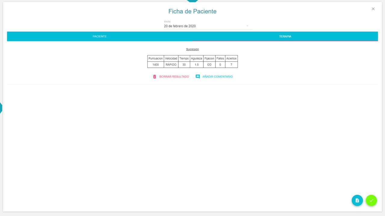
It is important to remember that to save the results, it is necessary to have previously selected the patient from the database.
Finally, within the test results, a button to Add Comment is shown so we can include any observation we wish regarding the exercised performed.
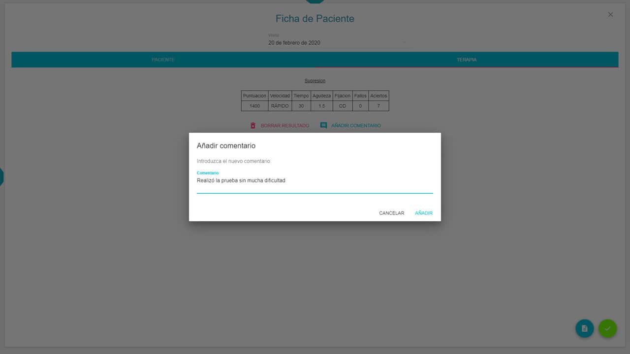
Once the comment is entered, it will appear in the file, along with the option to Delete that comment by pressing the X symbol.
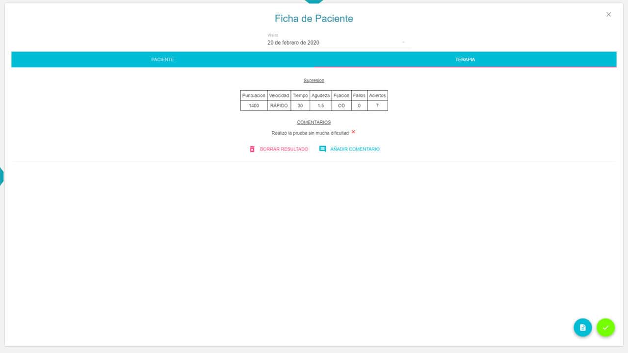
All recorded results also offer the option to Delete Result.
Use #
In general, the Block exercise can be used to treat suppression, for example, in patients with amblyopia, due to anisometropia or accommodative esotropia (when the eyes are well aligned*). It can also be useful for improving mild suppression and binocular coordination in patients with non-strabismic binocular disorders, for example, in convergence insufficiency (CI).
In the patient with CI, as they improve, we can ask them to move closer to the screen during the exercise to decrease the fixation distance, since at a shorter distance, it is more difficult to maintain binocular vision.
*Care must be taken not to lift suppression in patients with strabismic amblyopia; because if the angle of strabismus is not eliminated first, suppression is necessary to prevent diplopia and confusion. If we lift their suppression without aligning the eyes, each fovea will receive a different image (leading to confusion), and the same image will be stimulating non-corresponding retinal areas in each eye (diplopia). Once suppression is lifted, it is difficult to return to it, especially after childhood.
